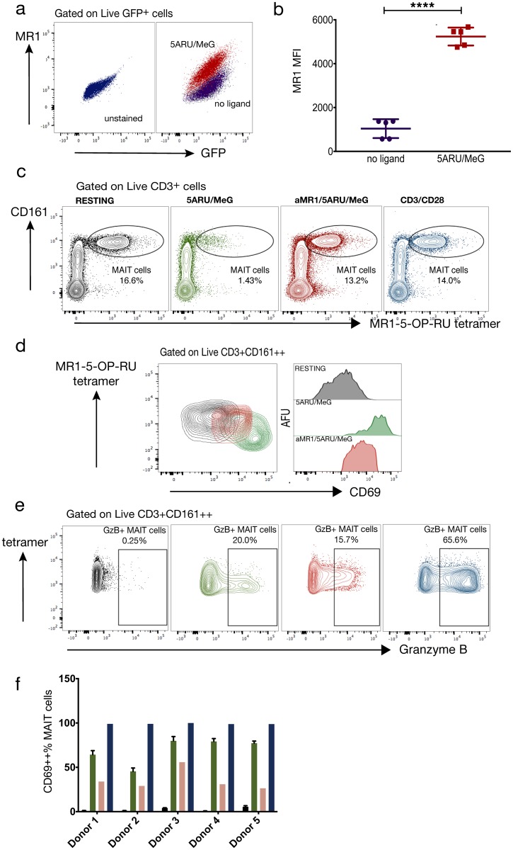
Fig 3. Functional studies.
5-Amino-6-(D-ribitylamino)uracil (5-A-RU) reacted with methylglyoxal (MeG) upregulates MR1 and activates human mucosal-associated invariant T (MAIT) cells. (a, b) 2 μM 5-A-RU•H2O and 50 μM MeG•H2O were directly incubated with the human C1R GFP MR1 expressing cell line for 15 hours. MR1 mean fluorescence intensity (MFI) was compared to resting C1R GFP by unpaired t-test. Graphed values are five technical replicates from two independent experiments; violet = no ligand, red = 2 μM 5ARU/50 μM MeG. **** p<0.001 (c) Human MAIT cells identified by flow cytometry as Live CD3+ MR1 tetramer+ CD161++ cells after 15 hours under the following conditions: black = no ligand, green = 2 μM 5ARU/50 μM MeG, red = αMR1/2 μM 5ARU/50 μM, blue = anti-CD3/CD28. Color code also applies to (d)-(f). (d) Contour plots and histograms representing MR1-dependent CD69 expression of MAIT cells in one human donor (e) Contour plots of MAIT cell granzyme B production under the same conditions in panel (c). (f) Mean and SD of MR1-dependent MAIT cell CD69 expression in five human donors. Mean and SD for resting and 5-A-RU/MeG conditions represent two technical replicates per condition. SD: standard deviation.

