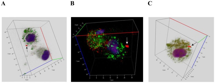
Fig 3. CD14 staining of alveolar macrophages with Mtb performed after ex vivo culture for 18 hours.
(A, B, C) Representative confocal fluorescent 3D images show alveolar macrophages stained by human CD14-specific (green signal) and Mtb Ag38-specific antibodies (red signal). Nuclei are stained by DAPI (blue signal). Alveolar macrophages were obtained from the resected lungs of patients 16 (A, B) and 18 (C). Black or white arrows indicate a single Mtb (A) and mycobacterial colonies (B, C) residing within alveolar macrophages.

