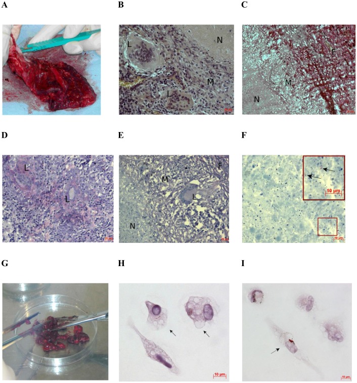
Fig 4. Histological examination of the resected lung from patient 7 shows the absence of cells with Mtb. Ex vivo analysis of alveolar macrophages disagrees.
(A) A surgically removed part of the lung. (B-F) Multiple TB lesions detected by histological analysis of tissue from the resected lung (A): N, caseous necrosis; M, a ring of macrophages; F, fibrous capsule; L, multinucleate Langhans giant cell. The scale bars are (B, D, E) 20 μm each, (C) 50 μm, and (F) 10 μm. (B, C, and E, F) Representative histological images show enclosed necrotizing granulomas stained by hematoxylin/eosin, a mixture of picric acid and fuchsin acid to detect collagen, and after the ZN method, respectively. (D) Multinucleate Langhans giant cells determined in an early non-necrotizing granuloma by ZN staining. (F) Scanty Mtb revealed in the caseous matter. Enlarged view of the part of this image with Mtb indicated by black arrows is shown in the upper right corner. (G) The tissue specimen of the resected lung (A) was cut into small pieces for producing alveolar macrophages. The diameter of the Petri dish is 10 cm. (H, I) Alveolar macrophages stained by the ZN method after ex vivo culture for 18 hours contain Mtb in isolation and as a colony, respectively. The black arrows point to alveolar macrophages with acid-fast Mtb. The scale bars are 10 μm each.

