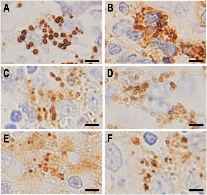
Fig 4. Higher magnification of small round bodies detected in the lymphatic sinus of sarcoid lymph nodes by IHC with each antibody.
A: IHC with PAB antibody after MT treatment, B: IHC with anti-IgG antibody, C: IHC with anti-IgA antibody, D: IHC with anti-IgM antibody, E: IHC with anti-C1q antibody, and F: IHC with anti-C3c antibody. Note that the dark brown-colored reaction products produced by each antibody are all located along the peripheral rim of the small round bodies. Scale bar: 5 μm.

