Fig. 2.
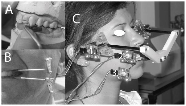
Results from dynamic stereometry are illustrated for one subject. Specifically, three-dimensional reconstructions of right and left temporomandibular joints from magnetic resonance images are shown in static superior-anterior views where the disc has been removed for better visualization of the ghosted images of the eminences in light grey over the condyles in shaded darker grey. Also shown superimposed over each condyle are the time-dependent positions of the centroid of the stress-field calculated at 5 ms intervals during jaw closing from laterotrusion (green dots) and during symmetric closing (red dots) as determined from combining the TMJ anatomy and jaw tracking data in three-dimensions. The location of the stress-field centroid for any given time-point was determined by finding the minimum condyle-fossa/eminence distance. The component variables of interest (x, aspect ratio; ΔD, distance of stress-field translation (mm); V, velocity of stress-field translation (mm/s); and Q, TMJ disc cartilage volume under the stress-field (mm3) were calculated for each joint and jaw closing movement at 5 ms intervals.

