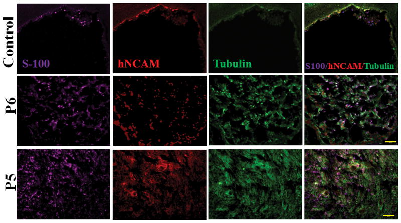
Figure 3.

Immunohistochemistry of regenerated nerves stained with S-100 (Schwann Cell Marker), hNCAM (human cell marker), and Tubulin (axon marker) showed that the p5 group clearly displayed stronger and more diffuse signals than the p6 group and control group. Scale bar: 100 μm.
