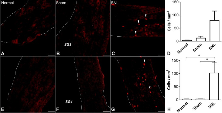
Fig. 8.
Effect of L5 spinal nerve ligation on infiltration of T cells in SG3 and SG4 on POD 7. A–D SG3 sections stained with an antibody to the T cell receptor (TCR) in normal (A), sham (B), and SNL (C) rats. The statistical analysis of TCR-positive cells/µm2 revealed that the differences between normal, sham, and SNL rats (D) did not quite reach significance (ANOVA, P = 0.0595). E–H SG4 sections stained for TCR in normal (E), sham (F), and SNL (G) rats showing that significantly more T cells were recruited in SNL SG4 than sham or normal rats (H; *P < 0.05, ANOVA with Tukey’s post-test). Dashed lines indicate ganglia borders. Arrowheads indicate individual T cells. (n = 4 male rats per group; scale bars, 50 µm).

