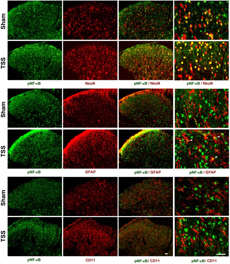
Fig. 2.
Expression of pNF-κB in the ipsilateral dorsal horn on day 3 after TSS and sham-operation. The cell markers and pNF-κB were visualized by double immunofluorescent staining. NeuN, neuronal marker; GFAP, astrocytic marker; CD11 (labeled by the antibody OX-42), microglial marker. Scale bars, 25 μm.

