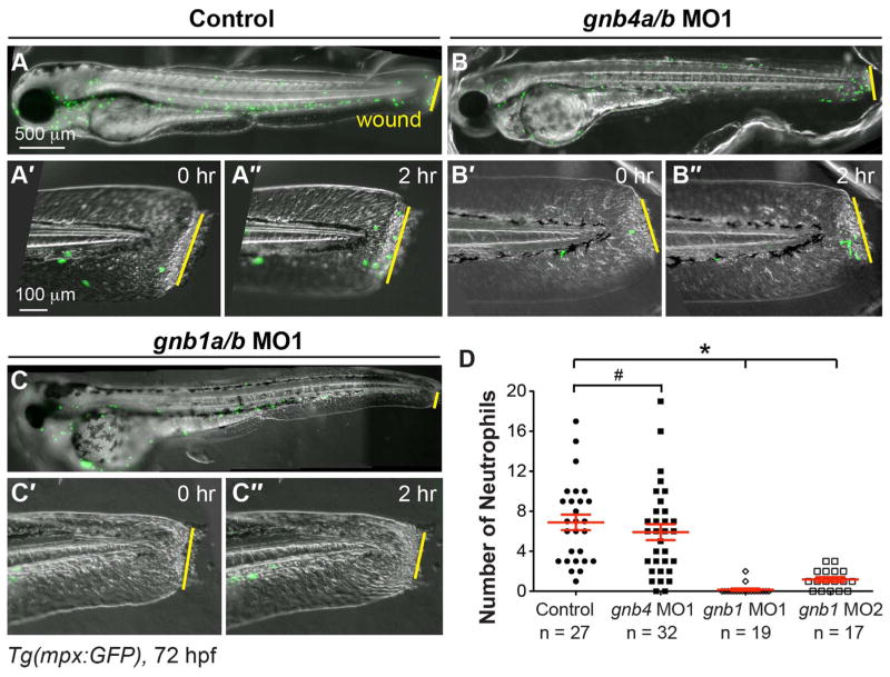
Fig. 3. Gβ1, but not Gβ4, is required for wound-induced neutrophil migration.
Epifluorescence time-lapse experiments tracking neutrophil migration for 2 h following wounding of the indicated 72 hpf-Tg(mpx:GFP) embryos. (A–C″) Overlay of epifluorescence and bright-field images of the whole embryo (A–C) and the tail region, immediately after wounding (0 h, A′–C′) and 2 h after wounding (2, A″–C″). Yellow lines: areas of wounding. (D) Number of neutrophils that reach the wound sites within 2 h of wounding (the number of embryos analyzed is indicated).

