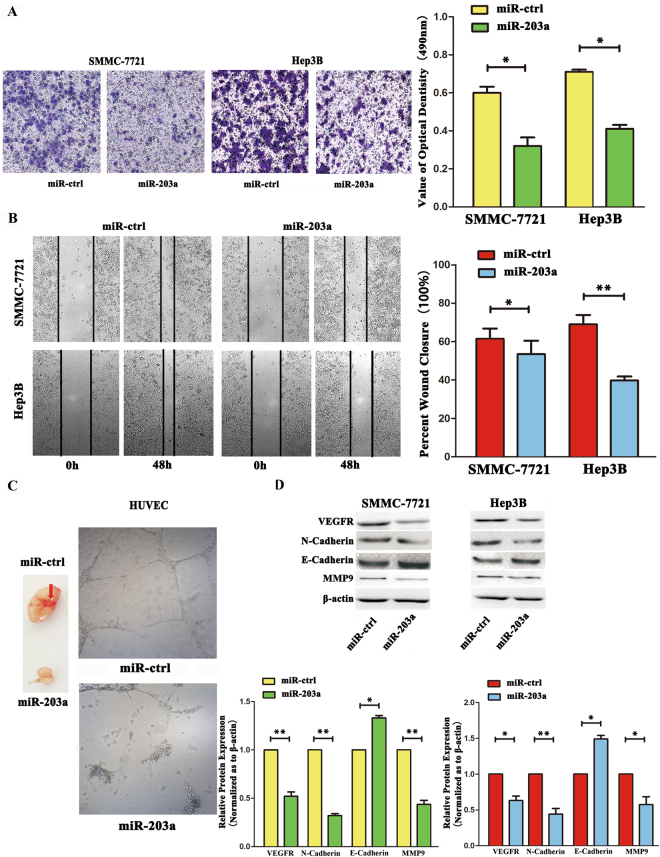
Figure 1.
Overexpression of miR-203a suppressed HCCs metastasis, invasion and angiogenesis. (A) Transwell analysis of SMMC-7721 and Hep3B cells after transfected with miR-203a and its ctrl, quantitative analysis of the invasion rates by solubilization of crystal violet and spectrophotometric reading at OD 490 nm. (B) Wound-healing assays of SMMC-7721 cells and Hep3B after treatment with miR-ctrl, miR-203a. The relative wound closure (100%) represents the metastasis capacity of SMMC-7721 and Hep3B cells. (C Left) Tube formation assay in HUVECs after transfected with miR-ctrl and miR-203a in vitro. (C Right) Angiogenesis assay in SMMC-7721 after transfected with miR-203a and its ctrl. (D) Effects of miR-203a overexpression on protein expression of EMT markers and related proteins by western blot. β-actin was detected as an internal control (*P < 0.05; **P < 0.01, Student’s t test).

