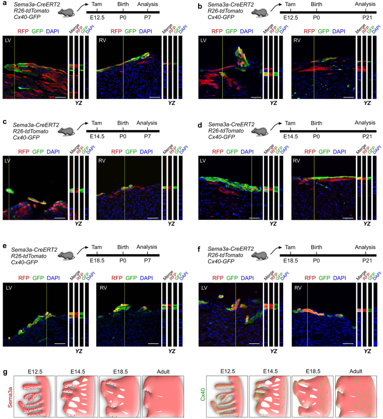
Figure 3.
Specialization of Sema3a+ cardiomyocytes into the conduction system in the developing heart. (a–f) Z-stack images of RFP and GFP immunostaining on Sema3a-CreERT2; R26-tdTomato; Cx40-GFP heart sections. Tamoxifen was administered at E12.5 (a,b), E14.5 (c,d) and E18.5 (e,f). The hearts were collected at P7 and P21 for each group. YZ indicates signals from the dotted lines on the Z-stack images. Scale bars, 50 µm. Each image is representative of 5 individual samples. (g) Schematic figure showing Sema3a+ cells (red) and Cx40+ cells (green) in the developing and adult heart.

