Figure 4.
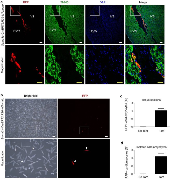
Quantification of the numbers of Sema3a+ cardiomyocytes in the adult heart. (a) Immunostaining for RFP and TNNI3 on adult heart sections. IVS, interventricular septum; RVW, right ventricular wall. Scale bars: white, 100 µm; yellow, 50 µm. (b) Image of isolated cardiomyocytes from the adult Sema3a CreERT2; R26-tdtomato heart showed Sema3a+ cardiomyocytes. (c) Quantification of the percentages of RFP+ cardiomyocytes in the heart sections. (d) Quantification of the percentages of RFP+ cells from among isolated cardiomyocytes. Each image is representative of 5 individual samples.

