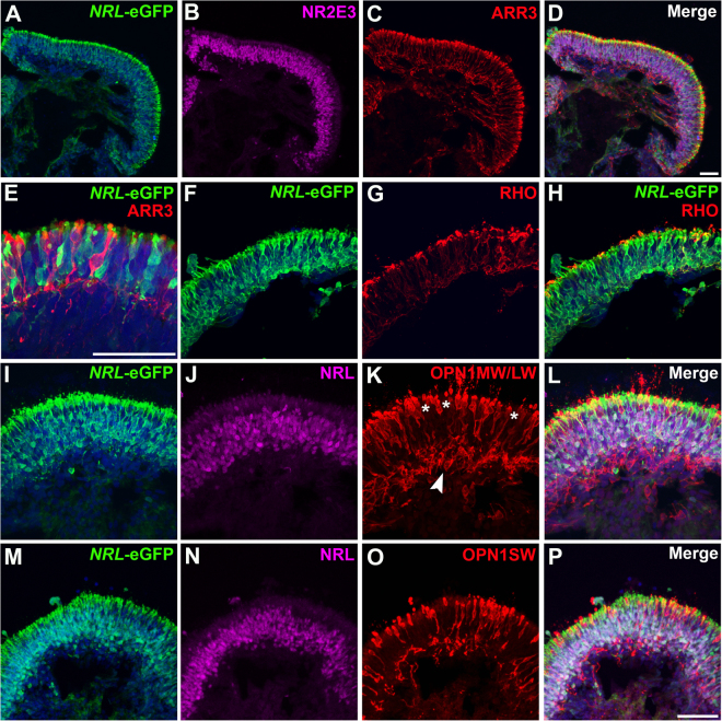
Figure 5.
eGFP labeling of rods in mid-stage NRL+/eGFP OVs. To examine mid-stages of rod differentiation in NRL+/eGFP OVs, d145 OVs were analyzed. (A,B) A large increase in rod production was observed by d145, as shown by co-expression of NRL-eGFP+ (A) and NR2E3 (B). (C,D) Numerous ARR3+ cones were also present at this time (C; merged image in D). (E) Higher magnification image of showing non-overlapping NRL-eGFP and ARR3 labeling in rods and cones. (F–H) Many NRL-eGFP+ rods (F) also expressed rhodopsin (RHO) (G; merged image in H) at this stage of differentiation. (I–P) Analysis of NRL-eGFP and NRL expression relative to red/green cone opsin (OPN1MW/LW; I–L) or blue cone opsin (OPN1SW; M–P) expression showed complete segregation of all cone opsins from NRL-eGFP+/NRL+ cells. Note the predominant localization of cone opsin-expressing cells along the outer (apical) portion of OVs (asterisks in K), although some cones were also found beneath the organized photoreceptor outer nuclear layer (arrowhead in K). Scale bars = 50 µm.

