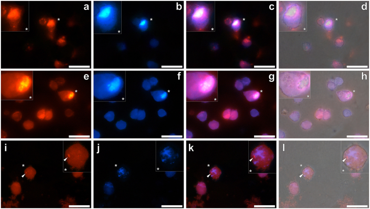
Figure 3.
Representative fluorescence microscopy of whole THP-1 cells cultured for 24 h in R10 medium in the presence of a simulated vaccine formulation. Vaccine treatments were prepared via the addition of 8 μM Aβ42 and 25 or 12.5 μg/mL ABA to complete R10 medium. Following 21 h incubation at 37 °C, 20 μM ThT and 100 μM lumogallion was added to the respective treatments and incubated for a further 3 h, prior to their 1:1 addition to THP-1 cells. Cells co-cultured for 24 h with simulated vaccine treatments contained 4 μM Aβ42 and (a–d) 12.5 μg/mL Alhydrogel® (Brenntag), (e–h) 12.5 μg/mL Imject Alum™ (Pierce, Thermo Scientific) or (i–l) 6.25 μg/mL Adju-Phos® (Brenntag). Cells were fixed in 4% w/v PFA, washed in 50 mM PIPES buffer, pH 7.4 before mounting with Fluoromount™ (Sigma Aldrich) on poly-lysine coated slides. Lumogallion (orange) (a,e and i) and ThT (blue) fluorescence (b,f and j) is depicted (Olympus single bandpass U-WMBV2 filter cube) with magnified inserts denoted (*). Merged lumogallion and ThT channels (c,g and k), combined with the light overlay (d,h and l) are also depicted. Arrows highlight discreet adjuvant particles. Magnification X 1000, scale bars: 20 μm.

