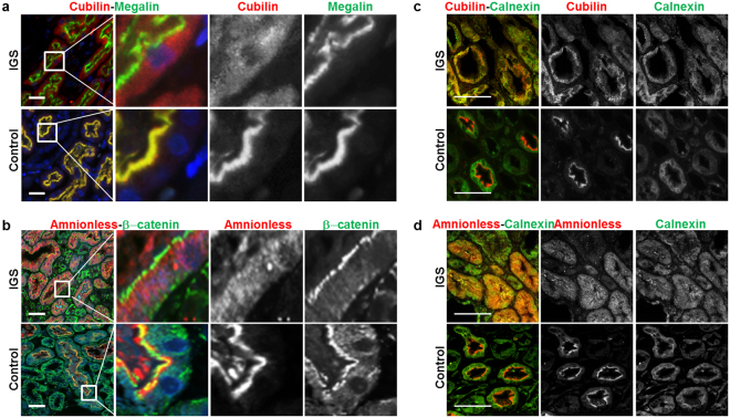
Figure 4.
Expression of cubilin and amnionless in human kidney cortex. Multi-labeling immunofluorescence staining for cubilin (red) and megalin (green) (a) or amnionless (red) and β-catenin (green) (b) in deparaffinized-embedded renal sections from the IGS patient and from a control subject (9-year-old boy with minimal-change nephrotic syndrome). Megalin is a multi-ligand binding receptor expressed in the apical membrane of tubular cells. β-catenin is expressed in the apical membrane of proximal tubular cell34. (c and d) While cubilin and amnionless were localized at the apical surface of proximal tubular cells in the control patient, those in the IGS patient showed abnormal intracellular localization. (Scale bar: 50 µm) Multi-labeling immunofluorescence staining for calnexin (green) and cubilin (red, (c)) or amnionless (red, (d)) in deparaffinized-embedded renal sections from the IGS patient and from a control subject. Cubilin and amnionless in the IGS patient were localized with calnexin. (Scale bar: 50 µm).

