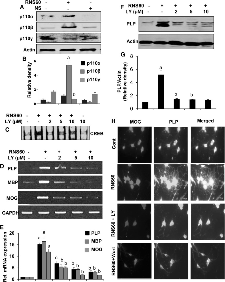
Fig. 7.
Role of PI3K in RNS60-mediated activation of CREB and expression of myelin-specific genes in primary mouse oligodendrocytes. Cells were treated with RNS60 for 30 min under serum free conditions followed by monitoring the levels of p110α, p110β and p110γ in membrane fractions by western blot (a). Bands were scanned and values are presented as relative density (b). Results are means ± SD of three different experiments. a p < 0.001 vs. control. Cells preincubated with different concentration of LY294002 (LY) for 30 min were treated with 10% v/v RNS60 under serum free condition. After 30 min of stimulation, activation of CREB was monitored by EMSA (c). After 6 h of stimulation, the mRNA expression of PLP, MBP and MOG was monitored by semi-quantitative RT-PCR (d) and real-time PCR (e). Results are means ± SD of three different experiments. a p < 0.001 vs. control; b p < 0.001 vs. control-RNS60; c p < 0.01 vs. control-RNS60. After 18 h of stimulation, the protein level of PLP was examined by western blot (f). Bands were scanned and values (PLP/Actin) are presented as relative to control (g). Results are means ± SD of three different experiments. a p < 0.001 vs. control; b p < 0.001 vs. control-RNS60; c p < 0.01 vs. control-RNS60. h Oligodendrocytes pre-incubated with either LY or wortmannin (wort) for 30 min were treated with 10% v/v RNS60 under serum free condition. After 18 h of treatment, cells were double-labeled with antibodies against MOG and PLP (g). Figures are representative of three independent experiments

