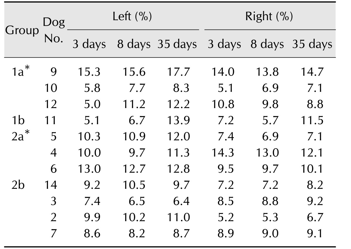
Table 3. Ventricular size at assessed times after middle cerebral artery occlusion in 11 dogs with infarcts.
Group 1a, dogs with unilateral cerebrocortical lesions; Group 1b, dogs with bilateral cerebrocortical lesions; Group 2a, dogs having unilateral lesions without cerebrocortical lesions; Group 2b, dogs having bilateral lesions without cerebrocortical lesions. *Groups 1a and 2a had unilateral lesions on the left side.

