. 2018 Jan 23;19(1):79–87. doi: 10.4142/jvs.2018.19.1.79

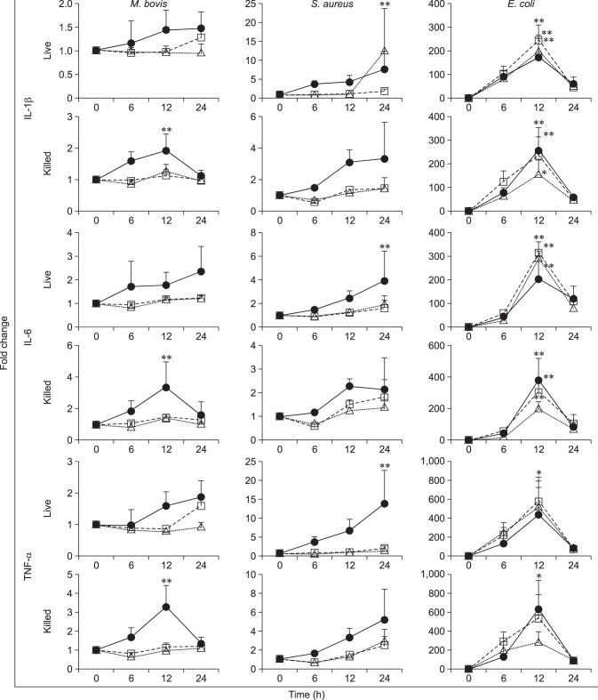
Fig. 2. Expressions of cytokine mRNA in bovine mammary epithelial cells (bMECs) stimulated with Mycoplasma bovis, Staphylococcus aureus, or Escherichia coli; bMECs were incubated with M. bovis, S. aureus, or E. coli, live or heat-killed, at multiplicities of infection of 10 (△), 100 (□), and 1,000 (●) for 6, 12, and 24 h. Expressions of interleukin (IL)-1β, IL-6, and tumor necrosis factor alpha (TNF-α) mRNA determined by real-time polymerase chain reaction and expressed as fold increases, as described in Materials and Methods section. The data are expressed as mean ± SE values from five independent experiments. Significant differences (*p<0.05 or **p<0.01) compared with unstimulated bMECs.

Fig. 2