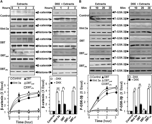
FIGURE 6.
Danggui Buxue Tang and DBTΔfa activate β-catenin translocation and GSK phosphorylations. Serum free osteoblasts were pre-treated with fresh medium, or 100 ng/mL of DKK, for 3 h before the application of 1.0 mg/mL of DBT for indicated time. (A) Beta-catenin (∼95 kDa) was detected by immunoblot analysis using specific antibodies, Histone-1 (∼30 kDa) served as an internal control. (B) Phosphorylation of GSK-3β (∼47 kDa) was detected by immunoblot analysis using specific antibodies, total GSK-3β (∼47 kDa) served as an internal control. Quantification of phospho-protein expression was calculated by a densitometer. Wnt3a (200 ng/mL) served as positive control. Values were expressed as the ratio to basal reading where the time zero (without treatment) equaled to 1, values were expressed as Mean ± SEM, where n = 3. ∗∗∗p < 0.001, ∗∗p < 0.01, and ∗p < 0.05 as compared to the control.

