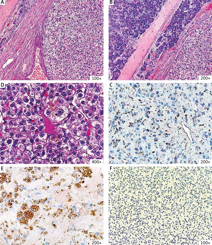
Fig. 2.
The histopathological images of the tumour. A–C) In the HE stain the tumour is composed of atypical neoplastic cells that form structures resembling the liver tissue. The accumulation of bile can be seen in C. D, E) The immunohistochemical stains reveal positivity for AFP and glypican-3 and negativity for chromogranin

