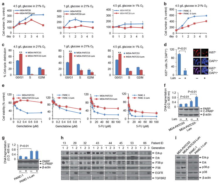
Figure 2.
Extracellular lumican induces phenotypic and signalling changes in PDAC cells. (a, b) MDA-PATC53 and PANC-1 (purchased from ATCC) cells were cultured with or without lumican (Lum; 2 μg/ml) in different culture conditions and cell number was counted. (c) Cell-cycle distribution was quantified by flow cytometry in MDA-PATC53 and MDA-PATC53-Lum cells in various culture conditions. **P<0.01. Parental MDA-PATC53 or PANC-1 cells were split to two parts, one was continuously exposed to 0.2 μg/ml lumican in culture medium around 30–90 days, named as MDA-PATC53-Lum or PANC-1-Lum cells; another was regularly cultured without lumican within the same period, serves as a control of MDA-PATC53-Lum or PANC-1-Lum cells. Cell DNA fingerprinting test at the Characterized Cell Line Core Facility of the MD Anderson Cancer Center (case number: CCLCF-YK-1421) verified the parental cells and lumican treated cells have the same DNA pattern and exactly match to primary cell line ID in our cell line database, suggesting they come from the same source and have the same basic genetic makeup. (d) Immunofluorescent staining showed Ki67-positive MDA-PATC53 cells after treatment with lumican for 3 days. Anti-Ki67 (sc-15402) was purchased from Santa Cruz Biotechnology. (e) MDA-PATC53, MDA-PATC53-Lum, PANC-1 and PANC-1-Lum cells were treated with gemcitabine or 5-fluorouracil (5-FU) for 72 h, and cell viability was measured by MTT assay. (f, g) Indicated cells were treated with lumican (2 μg/ml) for 24 h, cell lysates were subjected to a cell death detection ELISA kit (top) and western blot analysis (bottom). (h, i) Western blot results showed the expression of indicated molecules in PDX tumour tissues from different generations (h), and in MDA-PATC53, MDA-PATC53-Lum, PANC-1 and PANC-1-Lum cells (i). Anti- phospho-p44/42 MAPK (ERK1/2) (#9101), anti-p44/42 MAPK (ERK1/2) (#9102), anti-phospho-p38 MAPK (#9211) and anti-p38 MAPK (#9212) were purchased from Cell Signaling Technology. Anti-TGFBR2 (ab61213) was from Abcam.

