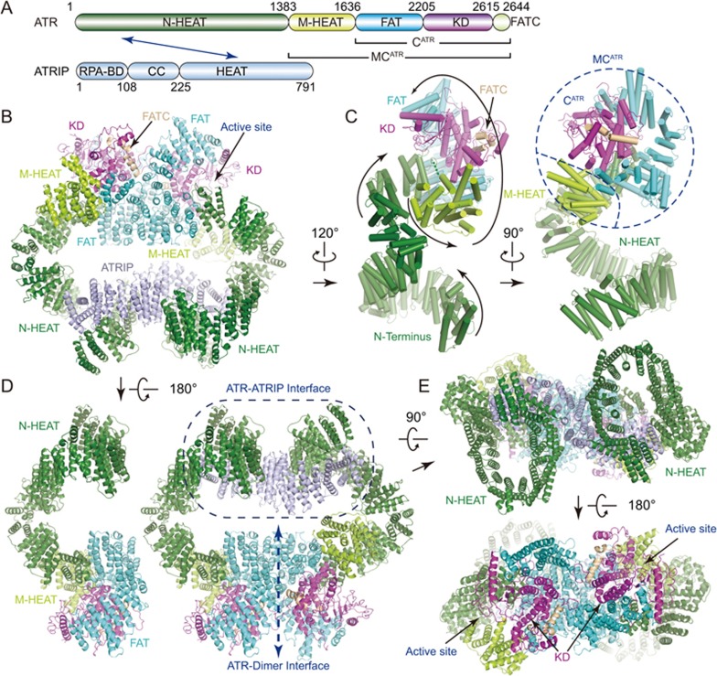
Figure 1.
Overall structure of ATR-ATRIP complex. (A) Color-coded domain structure of human ATR and ATRIP. The same color scheme is used in all structure figures if not otherwise specified. The domains involved in the interactions are indicated with arrow. (B-E) Ribbon representations of the ATR-ATRIP complex (B, E, and right panel of D) and ATR monomer structures (ATR-A in left panel of D, ATR-B in C) in different views. The topology of ATR is indicated with the peptide chain trace from the N- to C-terminus (C). RPA-BD, RPA-binding domain; CC, coiled coil; N-HEAT, N-terminal HEAT repeats; M-HEAT, middle HEAT repeats; FAT, FRAP, ATM, TRRAP domain; KD, kinase domain; FATC, FAT C-terminal domain.

