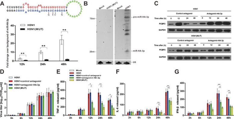
Figure 4.
Inhibition of miR-HA-3p reduces cytokine production in primary macrophages during H5N1 virus infection. (A) Levels of miR-HA-3p in primary macrophages infected with H5N1 or mutant H5N1 virus at different time points. Up: Schematic description of mutation site of H5N1 mutant. Down: Fold change of miR-HA-3p levels in primary macrophages detected by quantitative RT-PCR (qRT-PCR). (B) Northern blot analysis of miR-HA-3p using total RNAs extracted from primary macrophages infected with H5N1 or mutant H5N1 virus at 48 h post-infection. DIG-labeled LNA probe complementary to the sequence of miR-HA-3p was used. (C) PCBP2 protein levels in primary macrophages electroporated with control antagomir or miR-HA-3p antagomir prior to infection with H5N1 or mutant H5N1 viruses at different time points. (D) Viral titers of H5N1 or mutant H5N1 viruses in virus-infected primary macrophages determined by TCID50 assay using MDCK cells. (E-G) Levels of TNF-α (E), IL-6 (F) and IFN-β (G) in culture supernatants of macrophages infected with H5N1 or mutant H5N1 viruses plus different treatments. Data are presented as the mean ± SEM (n = 3). *P < 0.05. **P < 0.01.

