Figure 6.
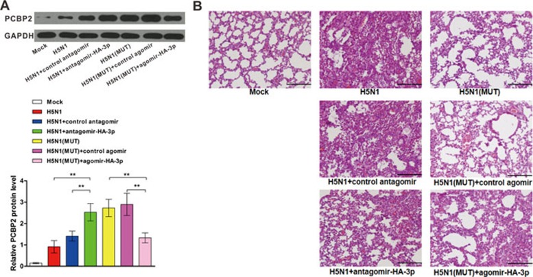
PCBP2 protein levels and histopathological effect on mouse lung tissues following H5N1 virus infection and different treatments. (A) Upper panel: representative western blot image of PCBP2 protein levels in mouse lungs on day 4 post-infection with H5N1 virus. (A) Lower panel: the quantitative analysis of the PCBP2 protein level in upper panel. Data are presented as the mean ± SEM (n = 3). **P < 0.01. (B) Representative images of histopathological effect of H5N1 infection on mouse lungs. Formalin-fixed, paraffin-embedded lung tissue sections from mice on day 4 post-infection were stained with hematoxylin and eosin. Note that lungs from H5N1-infected mice and H5N1-infected mice treated with control antagomir display acute neutrophil infiltration and necrosis of the bronchial and bronchiolar epithelium with considerable sloughing and disruption of the bronchial lining epithelium. In contrast, lungs from H5N1 virus-infected mice treated with antagomir-HA-3p or mutant H5N1 virus-infected mice are less severely damaged and display a mild to moderate neutrophil infiltration across the bronchial epithelium. Scale bars, 500 μm.

