
Fig. 6.
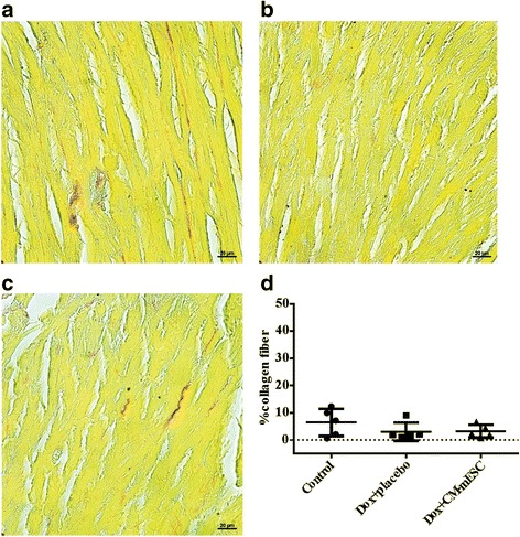
Histological analysis of the heart. a–c Representative images of histological sections stained with Sirius Red: a control group, b Dox + placebo group and c Dox + CM-mESC group after 45 days of cell therapy. d No changes in myocardial collagen fiber content. One-way ANOVA with a Bonferroni post test. Data shown as mean ± standard deviation; n = 5 for each group. Dox doxorubicin, CM-mESC cardiomyocytes derived from mouse embryonic stem cell