Fig. 3.

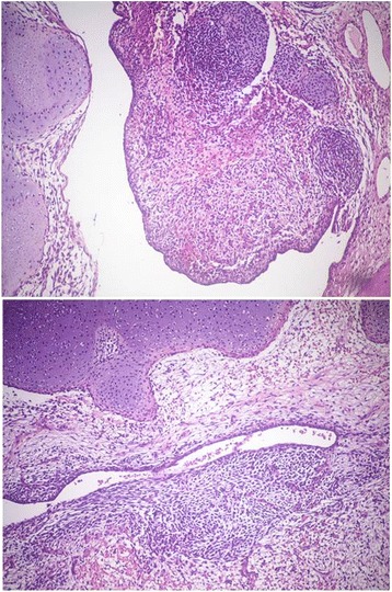
Medium-power view of the neoplasia, showing both the epithelial component and the undifferentiated spindle cell component, admixed with areas of cartilaginous differentiation (haematoxylin-eosin stain, 10X)

Medium-power view of the neoplasia, showing both the epithelial component and the undifferentiated spindle cell component, admixed with areas of cartilaginous differentiation (haematoxylin-eosin stain, 10X)