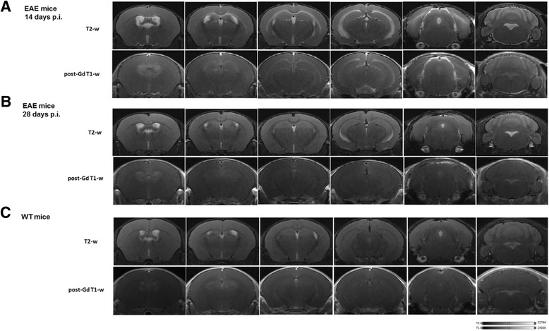
Fig. 2.

Magnetic resonance imaging (MRI) longitudinal study of a representative EAE mouse and healthy control. a Selected coronal MRI images acquired at 14 days p.i. showing focal lesions in T2-weighted and post-contrast T1-Gd-weighted images in different brain areas including hippocampus, corpus callosum, external capsule, and periventricular white matter. b Coronal T2 and T1-Gd MRI images acquired at 28 days p.i., representing the same brain coordinates of those shown in a. T2-enhanced regions are limited to the periventricular white matter and in the ventral aspect of hippocampus. EAE selected mouse exhibit 2.5 as clinical score at acute time point and 0 at late time point. c Selected coronal T2-weighted and post-contrast T1-Gd-weighted MRI images of a healthy control mouse
