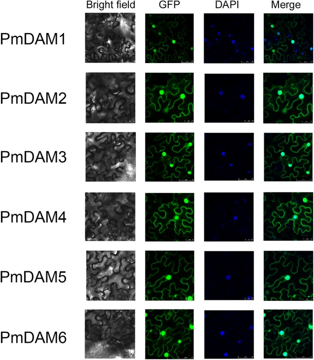
FIGURE 5.

Subcellular localization of six PmDAMs. To determine the exact position of PmDAMs within the cell, subcellular localization experiments were performed using leaf tissues of N. benthamiana. The green fluorescent showed protein position. The blue fluorescent presented the nuclei position. The merge pictures of PmDAMs were formed by the pictures of GFP and DAPI.
