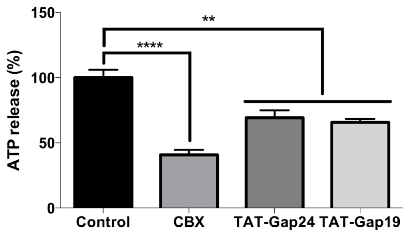
Figure 1. TAT-Gap24 and TAT-Gap19 block extracellular release of ATP.
Primary rat hepatocytes were isolated and cultivated as specified in “Materials and Methods”. 24 hours after cell seeding, cultured hepatocytes were exposed during 30 minutes to 20 µM TAT-Gap24, 20 µM TAT-Gap19, 50 µM carbenoxolone (CBX) or solvent control. Thereafter, the cells were exposed to HBSS-Hepes, considered as the baseline condition, and divalent-free buffer. After 2.5 minutes incubation at room temperature, ATP assay mix was added and luminescence was measured spectrophotometrically. ATP release in the divalent-free buffer were plotted and expressed as percentage of ATP release triggered by divalent-free medium. At least 6 wells per experiment were examined, with a total of 5 experiments (i.e. hepatocytes isolated from 5 different rats). Data are expressed as means ± SEM, with **p < 0.01 and ****p < 0.0001 compared to ATP release triggered by divalent-free medium.

