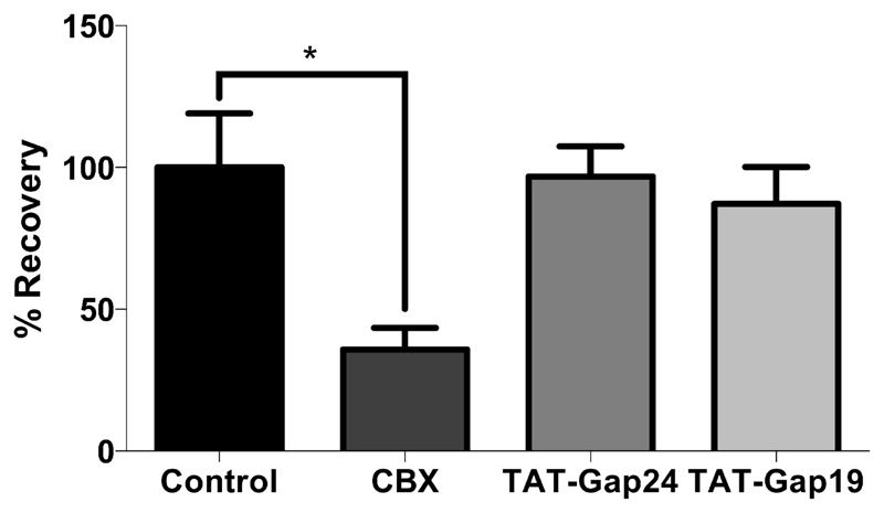
Figure 2. TAT-Gap24 and TAT-Gap19 do not affect gap junction activity.
Primary rat hepatocytes were isolated and cultivated as specified in “Materials and Methods”. 24 hours after cell seeding, cultured hepatocytes were exposed during 24 hours to 20 µM TAT-Gap24, 20 µM TAT-Gap19, 50 µM carbenoxolone (CBX) or solvent control. Subsequently, cells were subjected to FRAP analysis. After loading with 10 µM calcein-acetoxymethyl ester, fluorescence within a single cell was photobleached by 1 second spot exposure to 488 nm Argon laser. The dye influx from neighboring cells was recorded over the next 6 minutes. Fluorescence in the bleached cell was expressed as the percentage recovery relative to the prebleach level. At least 5 wells per cell culture dish were examined in each experiment, with a total of 5 experiments (i.e. hepatocytes isolated from 5 different rats). Data are expressed as means ± SEM, with *p < 0.05 compared to recovery in the solvent control.

