
Figure 1.
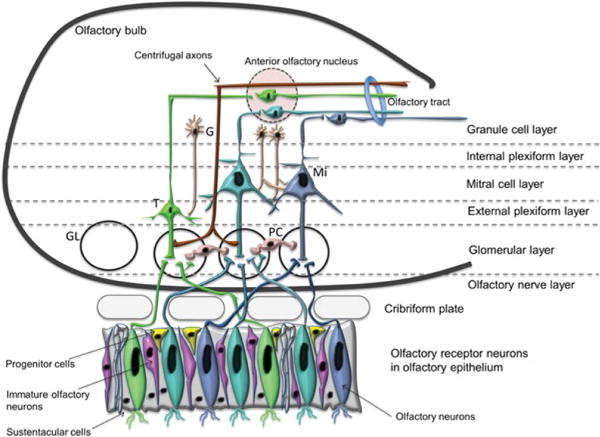
Schematic diagram of the olfactory neuroepithelium (OE) and the olfactory bulb (OB). The OE consists of cells at different stages of differentiation, including the proliferating progenitor cells (yellow color), the postmitotic immature olfactory neurons (pink color) and the olfactory sensory, OSN (also known as olfactory receptor neurons, ORN). Axons from the OSN pierce through the cribriform plate at the base of the skull to enter the OB, where they form the olfactory nerve layer. The OB, above the OE shows the laminar organization, the major cell types and the basic neuronal circuits. Interneurons shown are the granule cells (across different layers) and the periglomerular cells in the glomerular layer (GL). Efferent neurons of the olfactory bulb are tuffed and mitral cells.
Note: G: Granule cell; M: Mitral cell; T: Tufted cell; PC: Periglomerular cells