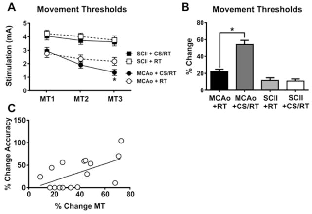
Figure 3. Minimum thresholds to evoke involuntary movement with cortical stimulation (MTs).
Cortical stimulation movement thresholds (MT1–3) were assessed on rehabilitation days 1, 10 and 19, respectively. A. Histogram comparing movement thresholds for animals given either middle cerebral artery occlusion (MCAo) or subcortical capsular ischemic injury (SCII). No significant differences were detected in movement thresholds between animals given SCII+RT and SCII+CS/RT. Animals given MCAo+CS/RT exhibited significantly lower movement thresholds relative to animals given MCAo+RT on MT3 (* p<0.05) whereas non-significant differences were detected between these groups at the other time points. B. Histogram comparing percent change in movement thresholds between MT1 and MT3. Animals given MCAo+CS/RT exhibited a significantly greater percent reduction in movement thresholds relative to animals given MCAo+RT (* p<0.05) whereas this difference was not observed in SCII animals. C. For animals given MCAo, a significant positive correlation was detected between percent change in reaching accuracy and percent change in movement thresholds (r= 0.57; p<0.05).

