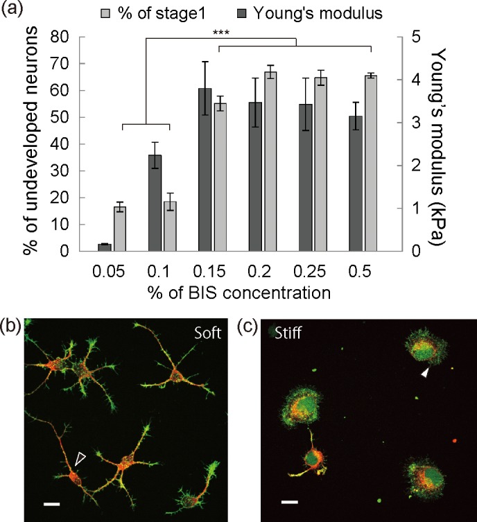
Fig 1. Substrate stiffness dependent neuritogenesis.

(a) Dark gray bars and light gray bars indicate the Young’s moduli of the gel substrates and neurons in stage 1 as a percentage of all the neurons on the gel substrates at 26 hours after plating, respectively. (b, c) Immunofluorescent images of representative neuronal morphologies on a soft gel substrate (b) and a stiff gel substrate (c) at 20 hours after plating. Red: β-III tubulin, green: F-actin.
