
Fig 4. Effects of cytochalasin D on neuritogenesis of neurons on gel substrates.
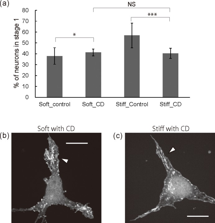
All neurons were exposed to 100 nM CD or DMSO for 20 hours. (a) Neurons in stage 1 as a percentage of all the neurons on the gel substrates with/without CD treatment. (n>300 cells per group, *p<0.05, ***p<0.001 by one-way ANOVA with Bonferroni post hoc test) (b, c) Fluorescent images of representative neurons on soft substrates (b) and on stiff substrates (c). All scale bars are 10 μm.