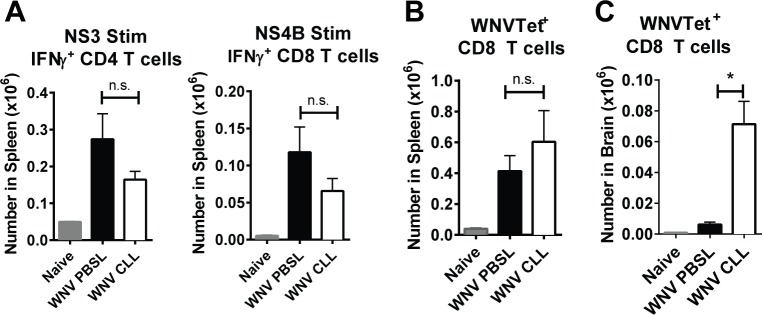
Fig 4. Generation of WNV-specific CD4 and CD8 T cells in WNV-infected splenic MΦ-depleted mice.
Mice were treated as in Fig 2A with CLL (open) or PBSL (black) and spleens and brains were harvested at day 8 p.i. (A) Splenocytes were stimulated with an NS3 peptide or an NS4b peptide and then analyzed for IFNγ expression within CD4 and CD8 T cells, respectively. (B) Splenocytes were stained with NS4b tetramers to enumerate WNV-specific (WNVTet+) CD8 T cells (C) Leukocytes from brains were stained with NS4b tetramers to enumerate WNVTet+ CD8 T cells. Statistics are shown for 1-way Anova plus Tukey’s post-test. P values: * p < 0.05, ** p <0.01, *** p<0.001.

