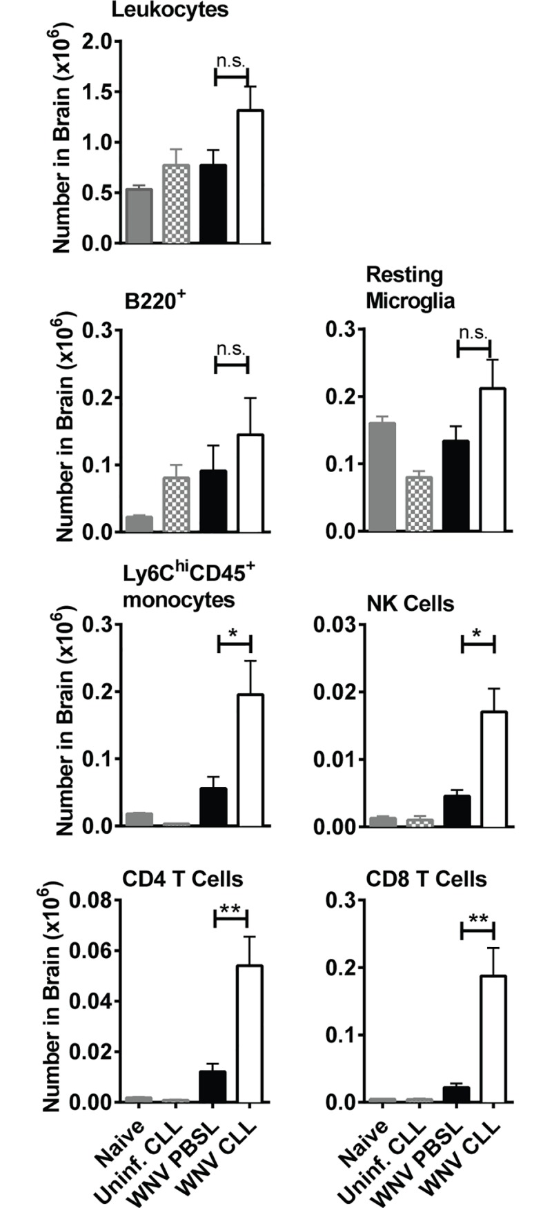
Fig 5. Cellular infiltration into the brains of WNV-infected splenic MΦ-depleted mice.

Brains were isolated from mice treated as in Fig 2A with either CLL (open) or PBSL (black), or from uninfected naïve mice (grey) or CLL treated mice (grey checked bar). Brains were harvested at day 8 post-WNV infection. Cell suspensions were stained with mAbs to surface markers and subsets quantified using flow cytometry as: CD45+ leukocytes, B220+ B cells, CD45lo CD11b+ Ly6C- Ly6G- resting microglial cells (52), CD45+ Ly6Chi CD11b+ Ly6G- monocytes, CD45+ NK1.1+ NK cells, CD45+ CD3+ CD4+ CD4 T cells, and CD45+ CD3+ CD8+ CD8 T cells. The results shown are the combined result of two independent experiments with similar results. Statistics shown are for two-tailed Student's t test, * p < 0.05, ** p <0.01, *** p<0.001.
