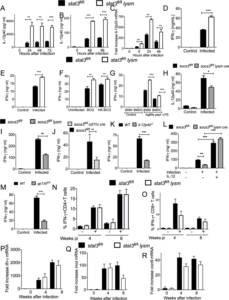
Fig 4. STAT3 in myeloid cells impairs IFN-γ secretion by mycobacteria-specific T cells in vitro.
The concentration of IL-12 p40 in supernatants from mycobacteria-infected stat3fl/fl lysm cre and stat3fl/fl BMDCs (A) or BMM (B) at different times after incubation were determined by ELISA. The mean IL-12p40 ± SEM pg/ ml from triplicate cultures is depicted. Differences with stat3fl/fl BMDCs are significant (**p<0.01, ***p<0.001 Student t test). Total RNA was extracted from stat3fl/fl lysm cre and stat3fl/fl BMDC cultures 24 h after M. tuberculosis infection. The mean Il-12p35 mRNA levels ± SEM levels measured by real time PCR are depicted (C) (**p<0.01 Student t test). Stat3fl/fl lysm cre and stat3fl/fl BMDC were infected with either BCG (D), M. tuberculosis (E) or stimulated with heat killed BCG (F) or with LPS and peptide 25 of Ag85b (G) washed and incubated 6 h after with p25-tg CD4+ naïve T cells (at a ratio of 4:1 BMDC) (G). The concentration of IFN-γ in the culture supernatants was measured by ELISA 72h after co-incubation. The mean IFN-γ ± SEM from triplicate cultures is depicted (***p<0.001 Student t test). The concentration of IL-12p40 in supernatants from mycobacteria-infected socs3fl/fl lysm cre and socs3fl/fl BMDCs was determined by ELISA (H). The mean IL-12p40 ± SEM ng/ ml from triplicate cultures is depicted (**p<0.01 and ***p<0.001 Student t test). Socs3fl/fl lysm cre (I), socs3fl/fl cd11 cre (J) and socs3fl/fl BMDC were infected with BCG and incubated 6 h after with p25-tg T cells. The concentration of IFN-γ in the supernatants was measured by ELISA 72h after co-incubation. The mean IFN-γ ± SEM from triplicate cultures is depicted (**p<0.01 Student t test). Il12p40-/- (K), gp130F/F (M) and WT BMDC were infected with BCG and co-incubated with p25-tg T cells as described. Mycobacterial-infected socs3fl/fl lysm cre and socs3fl/fl. were cultured in presence of recombinant IL-12p70 or left untreated (K) and co-incubated with p25Tg-T cells (L). The mean IFN-γ ± SEM in supernatants from triplicate cultures was measured by ELISA (**p<0.01, ***p<0.001 Student t test). The frequency of IFN-γ-secreting cells in PPD-stimulated pulmonary T cells from stat3fl/fl lysm cre and stat3fl/fl mice 4 and 8 weeks after infection with M. tuberculosis was analysed by ICS (N, O). The mean frequency of IFN-γ-secreting within CD4+ cells (N) and CD8+ (O) ± SEM is displayed (n = 5 per group). The levels of ifng (P), inos (Q)and cxcl9 (R) mRNA in the lungs of stat3fl/fl lysm cre and stat3fl/fl mice before and at the indicated time points after aerosol infection with M. tuberculosis were determined by real time PCR.

