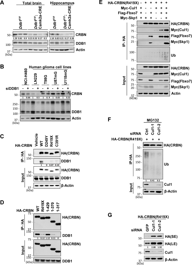
Fig 3. When dissociated from DDB1, CRBN and its ID-associated mutants are subjected to SCFFbxo7-mediated destruction.
Data are representative of experimental duplicates except for Fig 3A. (A) Western blot analysis for CRBN levels in protein extracts prepared from mouse brain (Ddb1F/F n = 3; Ddb1F/F;Camk2a-Cre n = 4) or hippocampus (Ddb1F/F n = 2; Ddb1F/F;Camk2a-Cre n = 3). Protein signals were measured by NIH Image J. Quantification of CRBN were standardized as the ratio of CRBN signals to the cognate actin signals, followed by normalization to the mean value of standardized CRBN signals in WT mouse brain or hippocampus. (B) Western blot analysis for CRBN levels in indicated cells transfected with siRNA for DDB1 or siRNA for GFP. siGFP, as a non-targeting control. Quantification of CRBN was normalized to the cognate actin signals. (C) Co-immunoprecipitation (co-IP) of HA-tagged WT or mutant CRBN with DDB1 in 293T cells. Whole-cell extracts from 293T cells over-expressing HA-CRBN constructs were immunoprecipitated with anti-HA affinity beads and analyzed by immunoblotting with indicated antibodies. Quantification of DDB1 pulled down by HA-CRBN in IP samples was normalized to the cognate DDB1 signals in input samples. (D) Identification of protein binding region of CRBN with DDB1 by immunoprecipitation of HA-tagged WT, R419X mutant or truncated CRBN with DDB1 after they were over-expressed in 293T cells. Quantification of DDB1 pulled down by HA-CRBN in IP samples was normalized to the cognate DDB1 signals in input samples. (E) Co-immunoprecipitation of CRBNR419X with Fbxo7, Skp1, Cul1 and ubiquitin in 293T cells over-expressing HA-CRBNR419X, Flag-Fbxo7, Myc-Skp1 and Myc-Cul1. (F) Ubiquitination assay for CRBNR419X by immunoprecipitation of CRBNR419X with ubiquitin in 293T cells. 293T cells over-expressing HA- CRBNR419X were transfected with siRNA for Cul1 or control and harvested for IP after treatment with MG132 (10 μM) for 6 hrs. Quantification of Ubiquitin pulled down by HA- CRBNR419X was normalized to β-actin. (G) Western blot analysis for CRBNR419X levels in 293T cells transfected with HA-CRBNR419X along with siRNA for Ctrl or Cul1. L.E., long exposure. S.E., short exposure. Quantification of CRBNR419X (L.E.) was normalized to β-actin.

