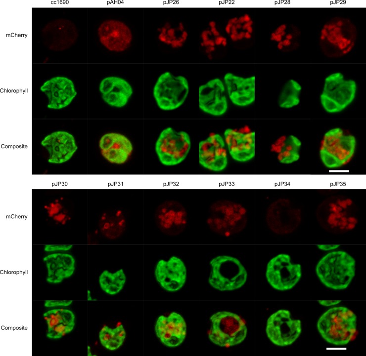
Fig 5. Live-cell fluorescence microscopy of the mCherry-expressing strains.
The mCherry signal from the non-secreting pAH04 construct is distributed in the cytosol, while the secreting pJP transformants present a mCherry signal in vesicles. Live cells were plated on agar pads and images were acquired 0.4 -μm apart in each channel in the z-axis. Then, images were stacked using the Fiji software Z projects function, generating the final images. An argon laser at 543 nm was used to excite mCherry, and a spectral detector set at approximately 610–650 nm was used to detect emitted fluorescence. For chlorophyll, we used a laser at 405 nm for excitation, and a spectral detector set at 680 nm. cc1690 –parental wild-type strain; pAH04 –construct without SP; pJP22 –construct with arylsulfatase 1 SP; pJP26 –construct with binding protein 1 SP; pJP28 –construct with carbonic anhydrase 1 SP; pJP29 –construct with ice-binding protein 1 SP; pJP30-35 –construct with in silico identified SP. All images were processed identically. Scale bar = 5 μm.

