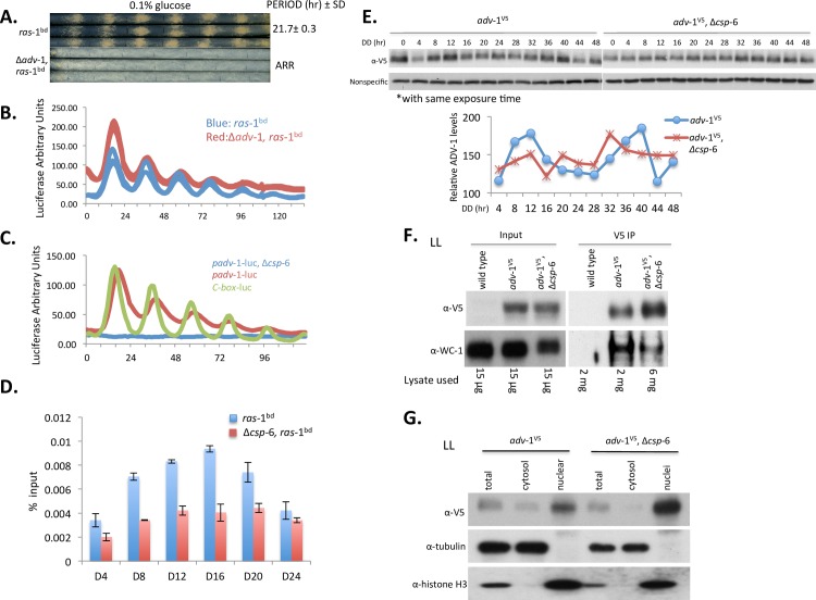
Fig 6. ADV-1 is a direct downstream target of CSP-6-WC-1 that regulates circadian output.
A: Race tube data showing loss of overt conidiation rhythmicity in Δadv-1; triplicate race tubes are shown. Period is reported in hours ± one standard deviation, ARR is arrhythmic. B: Luciferase traces of frq c-box-luc in the ras-1bd and Δadv-1, ras-1bd genetic backgrounds confirming that the clock functions normally in Δadv-1. C: Representative luciferase activity assays showing circadian rhythmicity of adv-1 promoter activity was significantly impaired in the Δcsp-6 mutant. D: ChIP assays were performed in a time course across a circadian cycle from DD4 to DD24 to show the recruitment of WC-2 to the adv-1 promoter in the ras-1bd and Δcsp-6, ras-1bd. Error bars reflect +/- 1SD, n = 3. E: Western blot analysis of protein expression of V5-tagged ADV-1 in WT and Δcsp-6 (Upper). The data shows protein ADV-1 is expressed rhythmically in the wild type strains but the rhythmicity was lost in Δcsp-6. Densitometric analysis of these data is shown in the lower panel. F: Co-IP assay demonstrating ADV-1 and WC-1 interaction using DSP crosslinking in constant LL. V5-tagged ADV-1 was co-immunoprecipitated with anti-V5 agarose beads and WC-1 was detected with anti-WC-1 antibody. To compensate for the reduced WC-1 present in Δcsp-6, 3 fold more lysate was used for the V5-IP in adv-1V5, Δcsp-6 as compared to adv-1V5; even so, reduced interaction between ADV-1 and WC-1 was detected in Δcsp-6 compared to WT. G: ADV-1 protein is enriched in nuclei. Subcellular fractions from adv-1V5 in WT and Δcsp-6 were analyzed with the indicated antibodies. Tubulin was used as a control to show nuclear fraction was not contaminated with cytosolic protein and histone H3 was used to show enrichment of the nuclear fraction.

