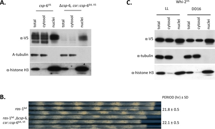
Fig 7. CSP-6 localizes inside nuclei in Neurospora.
A: Western blots show CSP-6 and two control proteins (tubulin and histone-H3) in total cell lysates (Total), cytoplasm (Cyto) and nuclear fractions of csp-6V5 and Δcsp-6, csr::csp-66A, V5. CSP-6 was detected in the nuclei; there was no tubulin signal in the nuclear fraction suggesting that isolated nuclei have little cytosolic contamination. B: Race tube assay showing Δcsp-6 ras-1bd; csr::csp-66A, V5 has normal overt circadian rhythmicity with the same period as wild type (ras-1bd); triplicate race tubes are shown. Period is reported in hours ± one standard deviation, ARR is arrhythmic. C: Subcellular distribution of WHI-2 was analyzed by Western blot with indicated antibodies. Cellular fractions were prepared by differential centrifugation by standard techniques (57), but the final nuclear pellet was resuspended in just 300 μl of buffer versus 40 ml for total and cytoplasmic fractions. In all cases the same volume of extract (10μl) was loaded on the gels, so estimates of the total amount of protein in each compartment must reflect both the amount seen on the gel and the total amount of extract prepared.

