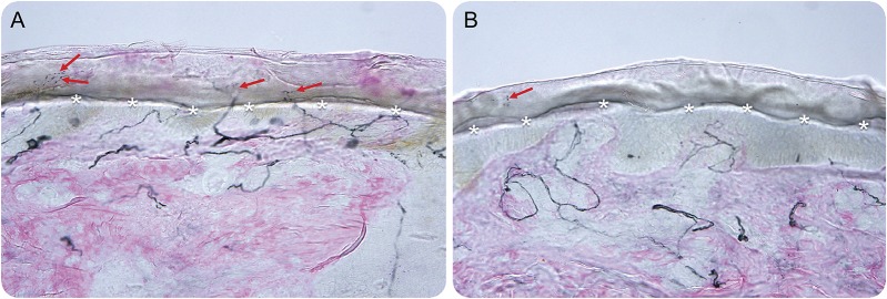
Figure. Skin biopsy findings diagnostic of a small-fiber neuropathy (SFN) in a patient with rheumatoid arthritis.
The patient had symptoms and clinical findings suggestive of SFN, including burning and allodynic pain in his feet, normal Achilles reflexes, preserved vibratory and joint position sensation, normal nerve conduction studies, and length-dependent diminution to pinprick up to the distal shins. Therefore, punch skin biopsy findings were taken from standardized sites at the right calf (A) and the right foot (B). The respective biopsy sites were 10 cm above the lateral malleolus in the calf and in the dorsum of the foot above the extensor digitorum brevis muscle. Panaxonal protein PGP 9.5 immunostaining was performed on the skin biopsy specimens. The intraepidermal nerve fiber density (IENFD) of unmyelinated nerves was quantified. The diagnosis of a SFN was ascertained when the IENFD was below the fifth percentile from normal controls. The asterisks correspond to the boundary between the epidermis and dermis. The arrows correspond to the nerve fibers. As shown in the figure, there is normal IENFD of unmyelinated nerves in a punch biopsy taken from the right calf. In contrast, the figure demonstrates that there is decreased IENFD of unmyelinated nerves in a punch biopsy taken from the right foot. This pattern in which there is a gradient of abnormally reduced IENFD at the distal site (figure) vs the proximal site (figure) is termed a length-dependent SFN, and is consistent with a pattern of primarily axonal degeneration.

