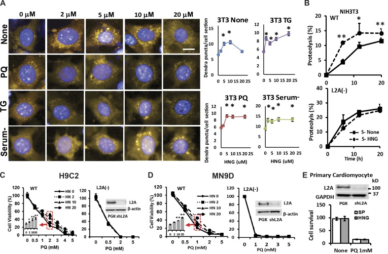
Figure 3.
HNG induces CMA, and induction of CMA contributes to the cytoprotective effects of HNG. (A) NIH3T3 cells stably transduced with lentivirus carrying a KFERQ-dendra reporter used to monitor CMA activity were incubated with indicated concentrations of HNG in the absence or presence of PQ, thapsigargin (TG), or serum deprivation (serum−). Left: Representative images. Bar, 10 µm. Right: Quantification of changes in the mean number of puncta per cell section quantified with high-content microscopy in n = 1,500 cells per condition. Differences are significant for *, P < 0.001. (B) Long-lived protein degradation in NIH3T3 cells WT (top) and knocked down for LAMP-2A (L2A−; bottom) supplemented or not supplemented with 10 µM HNG. n = 4. (C and D) Effects of different doses of HNG on cell viability in response to PQ on WT or L2A− H9C2 (C) and MN9D cells (D). (E) Effects of HNG on cell viability in response to PQ on L2A− primary cardiomyocytes. Differences with untreated cells were significant for *, P < 0.05; **, P < 0.001. Error bars show SEM.

