Figure 4.
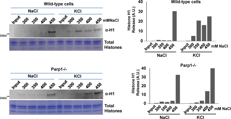
PARP1 mediates H1 release from chromatin in stimulated PMNs. Nuclei were isolated from WT (top) and Parp1−/− (bottom) in both NaCl-treated (left lanes) and KCl-stimulated (right lanes) PMNs and biochemically extracted with increasing salt concentrations (300–450 mM NaCl), as indicated above each lane. The extracted histone H1 fraction was detected by Western blotting with H1 antibody. As a loading control, the histones were isolated from the samples using an acid-extraction method, loaded in 18% SDS-PAGE gel, and stained with Coomassie blue. The intensity of the H1 Western signal was quantified using ImageJ software and shown to the right. Values on the y axis are arbitrary units.

