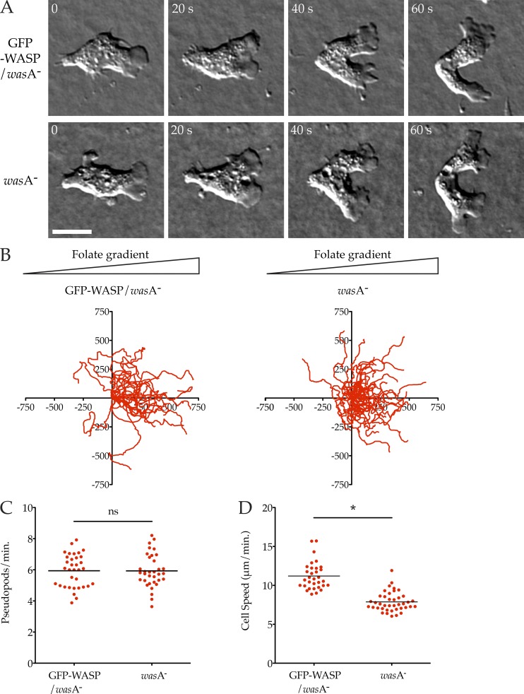
Figure 1.
WASP is not required for chemotaxis or pseudopod formation. (A) Normal pseudopods in WASP knockout cells. wasA− cells ± GFP-WASP were allowed to chemotax to folate under agarose and examined by DIC microscopy. A representative cell is shown. See also Video 1. Cells are shown at 20-s intervals. Bar, 5 µm. (B) Robust chemotaxis in WASP knockout cells. Cells (as in A) were tracked showing equivalent, strong bias in the direction of the chemoattractant (>20 cells/line from three independent assays; triangles indicate direction of the folate gradient; scale is in micrometers). (C) Rate of pseudopod formation. GFP-WASP/wasA− control and the wasA− mutant showed identical rates of pseudopod formation (5.94 vs. 5.93 pseudopods/min, respectively). (D) Diminished speed in WASP knockout cells. Speeds were derived from tracks in B, showing a decrease in wasA− cells compared with the GFP-WASP/wasA− control knockout (7.88 ± 0.20 vs. 11.20 ± 0.31 µm/min, mean ± SEM; P < 0.0001, unpaired Student’s t test).

