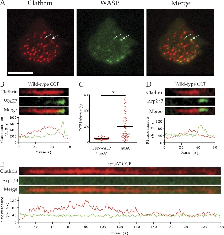
Figure 2.
Defects in clathrin-mediated endocytosis. (A) Colocalization of WASP and clathrin pits. wasA− cells were transfected with CLC-mRFP (clathrin) and GFP-WASP (giving a wild-type WASP phenotype) and imaged by TIRF. WASP colocalizes with a subset of clathrin pits at any time (white arrows). Bar, 10 µm. (B) Colocalization of clathrin and WASP at puncta. Wild-type cells were transfected with CLC-mRFP (clathrin) and GFP-WASP, and clathrin-mediated endocytosis was visualized using TIRF microscopy. Kymograph and fluorescence intensity plot demonstrate the dynamic localization of clathrin and WASP at clathrin pits (representative of >50 puncta from >20 cells). (C) Vesicle internalization during clathrin-mediated endocytosis. wasA− cells were transfected with CLC-mRFP alone or CLC-mRFP and GFP-WASP and visualized by TIRF. Clathrin punctum lifetime (time between appearance and disappearance from TIRF field of view) was measured from 50 pits/cell line over two independent experiments. Clathrin puncta in WASP knockout cells were far longer lived than in GFP-WASP/wasA− controls (197.5 ± 22.8 vs. 44.2 ± 1.7 s, mean ± SEM); P < 0.0001, unpaired Student’s t test). (D and E) Recruitment of Arp2/3 complex. Cells were transfected with CLC-mRFP (clathrin) and GFP-ArpC4 (Arp2/3 complex), and clathrin-mediated endocytosis was visualized using TIRF microscopy. Kymographs and accompanying fluorescence intensity plots demonstrate the dynamic localization of clathrin and the Arp2/3 complex at clathrin pits (representative of >50 puncta from >20 cells). (D) In wild-type cells, recruitment of the Arp2/3 complex to clathrin pits coincides with internalization. (E) In the wasA− mutant, many clathrin pits fail to recruit the Arp2/3 complex and persist on the plasma membrane for hundreds of seconds.

