Figure 1.
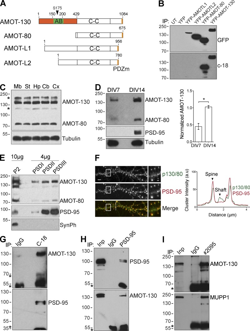
Distribution of AMOT isoforms in the CNS. (A) Schematic of structural domains in the four major AMOT isoforms. The location of the actin-binding (AB) region, the N-terminal region (orange), coiled-coil (C-C) domain, and the PDZ-binding motif (PDZm) are indicated. The arrowhead denotes the conserved serine (S-175) within the actin-binding domain. (B) Lysates from HEK293T cells transfected as indicated were immunoprecipitated (IP) and probed with anti-GFP antibody (top). Membranes were reprobed with C-18 antibody (bottom). UT, untranslated. (C) Expression of AMOT in the indicated brain regions. Mb, midbrain; St, striatum; Hp, hippocampus; Cb, cerebellum; Cx, cortex. Asterisks indicate unspecific bands. (D) Clarified lysates from 7- and 14-DIV hippocampal neuron cultures analyzed by immunoblotting with indicated antibodies. The graph to the right shows quantification of AMOT-130 protein normalized to tubulin. Error bars represent means ± SD. *, P < 0.05; Student’s t test. (E) Protein isolated from crude synaptosomal fraction (P2) and enriched PSD fractions were analyzed by immunoblotting. SynPh, synaptophysin. (F) Double immunostaining of PSD-95 (red) and AMOT-130/80 (green) in hippocampal neurons. Bars: (main images) 5 µm; (insets) 1.25 µm. Right: intensity profile along white line across the dendrite. (G–I) Coimmunoprecipitation with IgG or antibody recognizing endogenous AMOT-130/80 (C-18), PSD-95 (H), or AMOT-130 (2095; I), which does not recognize AMOT-80, from hippocampal lysates. Bound complexes were analyzed by immunoblotting (top) and reprobed (bottom) with the indicated antibodies. Asterisks denote IgG heavy chains. Molecular masses are given in kilodaltons.

