Figure 5.
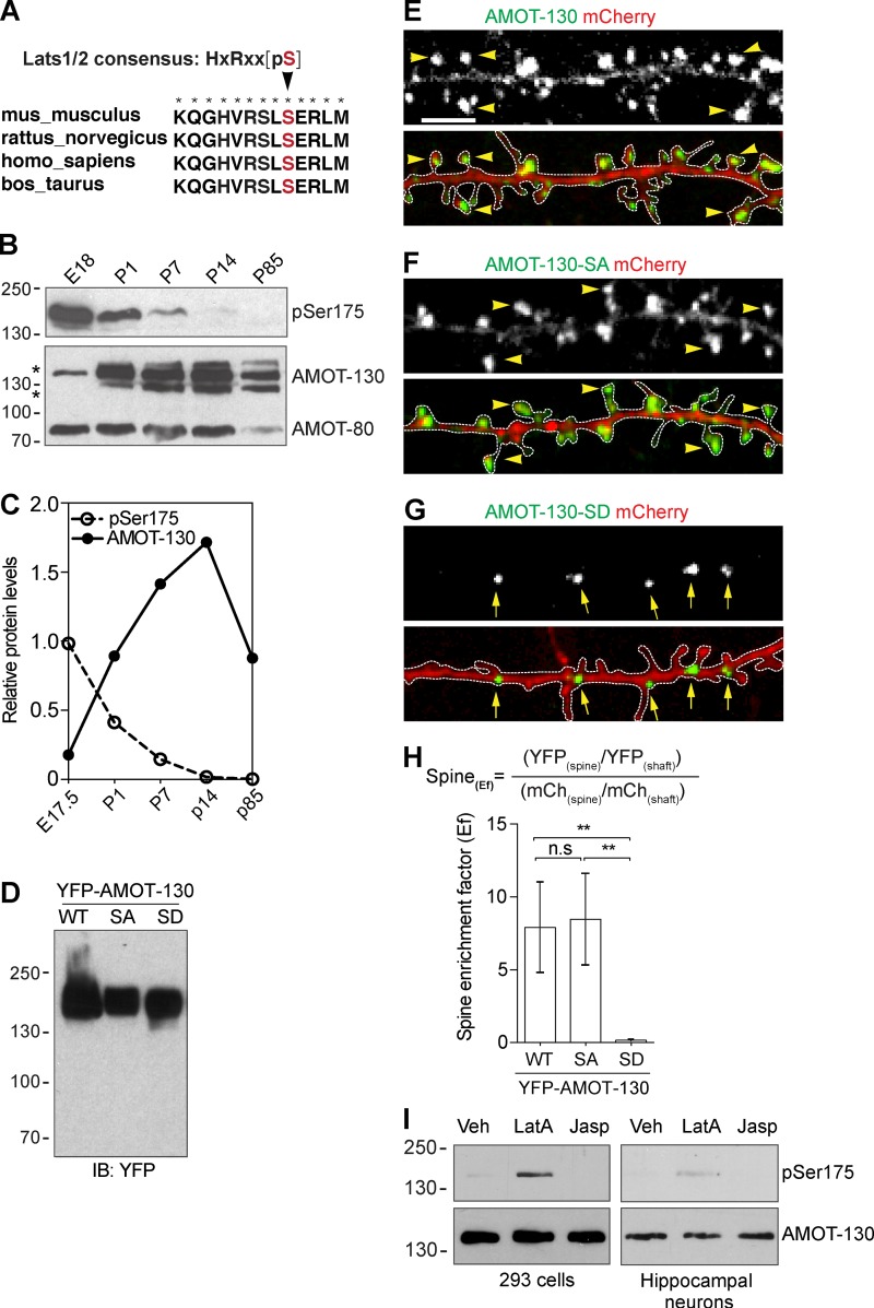
Phosphorylation at S-175 excludes AMOT-130 from spines. (A) Sequence alignment of AMOT-130. The arrowhead pointing to Ser175 (red) within the actin-binding domain of AMOT-130 is the consensus phosphogroup acceptor residue for the Lats1/2 kinases. Asterisks indicate fully conserved residues. (B) AMOT expression in the rat brain at the indicated stages analyzed by immunoblotting with antibodies against Ser175-phosphorylated AMOT-130 (pSer175) and total AMOT-130/80 protein. (C) Quantification of the relative expression of pSer175 and AMOT-130 protein in the rat brain. (D) Expression levels of YFP–AMOT-130–WT, YFP–AMOT-130–SA, and YFP–AMOT-130–SD in HEK293T cells. IB, immunoblot. (E–G) Neurons expressing mCherry together with YFP–AMOT-130–WT, YFP–AMOT-130–SA, or YFP–AMOT-130–SD. Panels show dendritic sections outlined by dashed lines with punctates in spines (arrowheads) or along dendritic shafts (arrows). Bar, 5 µm. (H) Spine enrichment of AMOT-130 by calculating the Ef. Error bars represent means ± SD. **, P < 0.01; one-way ANOVA with Tukey’s post hoc test. (I) HEK293T cells or 10-DIV hippocampal neurons were treated with Vehicle (Veh), LatA (2 µM), or jasplakinolide (2 µM) for 1 h and analyzed by immunoblotting as indicated. Molecular masses are given in kilodaltons.

