Figure 2.
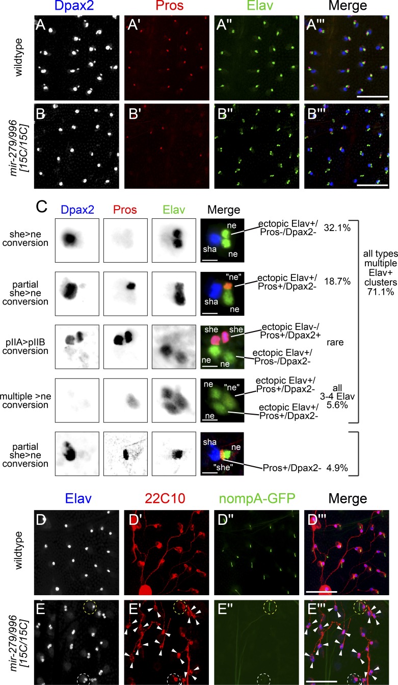
Profound sensory organ cell fate defects in mir-279/996 mutants. Shown are pupal nota at >28 h APF stained for cell-specific markers. (A and B) Wild-type (A) and mir-279/996[15C/15C] (B) nota stained for DPax2 (large shaft and small sheath nuclei), Pros (sheath nuclei), and Elav (neuronal nuclei) reveal a strong sheath-to-neuron transformation in the mutant, largely without affecting outer cell fates. Bars, 50 µM. (C) Higher magnification of mir-279/996[15C/15C] mutant sensory organs organized into characteristic classes of aberrant expression of cell-specific markers. In general, these represent conversion of non-neural cell types (especially sheath cells) into neuronal fate. Quantification of their frequency is provided at right, and their interpretation with respect to the canonical sensory lineage (see Fig. 1 A) is provided at left (wt, n = 792; mir-279/996[15C/15C], n = 593; see also Fig. 7 G for further phenotypic analysis). Bars, 10 µM. (D and E) Staining for the terminal markers of the neuron (22C10) and the sheath cell (nompA-GFP) in wild type (D) and mir-279/996[15C/15C] (E) indicates that mutant sensory organs differentiate as neurons and fail to elaborate the dendritic cap of the sheath. Dotted white circle in E′ highlights an exceptional case in which an ectopic Elav+ neuron is not surrounded by 22C10 reactivity; however, in the vast majority of preparations, the ectopic Elav+ cells express equivalent amounts of 22C10. Dotted yellow circle in E′ highlights a mutant sensory organ that elaborates a nompA-GFP sheath cell dendritic cap. Bars, 50 µM.

