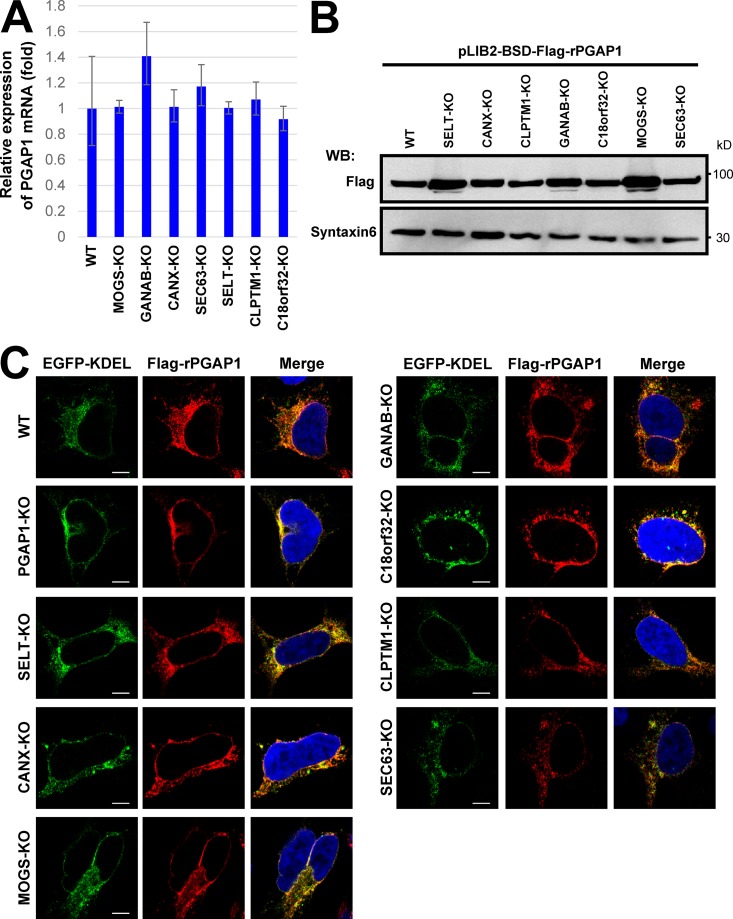
Figure 3.
Expression, protein stability, and localization of PGAP1 were not changed in gene KO cell lines. (A) Quantitative PCR analysis of PGAP1 mRNA levels in WT HEK293FF6, MOGS-KO, GANAB-KO, CANX-KO, SEC63-KO, SELT-KO, CLPTM1-KO, and C18orf32-KO cells. GAPDH values were used to normalize the data. The bars represent RQ (relative quantification) values ± RQmax and RQmin (error bars) of triplicate samples. (B) Cells stably expressing Flag-tagged rat PGAP1 (Flag-rPGAP1) were lysed and analyzed by Western blotting (WB). Proteins were detected with anti-Flag antibodies. Syntaxin 6 was used as the loading control. (C) Cells stably expressing Flag-tagged rat PGAP1 were transfected with GFP-KDEL and immunostained with an anti-Flag antibody. Images were collected using confocal microscopy. DAPI staining was shown as blue in merged images. Bars, 5 µm.

